

티스토리 뷰
[Cell culture] Stampwell V - 3D cell imaging [GRE-MOU-V300, GRE-MOU-V500]_Idylle - 코아사이언스
코피디 2024. 7. 11. 14:39Stampwell V - 3D cell imaging
Mold agarose wells to parallelize spheroid and organoid imaging
Cat.# Shape
GRE-MOU-V300 V300
GRE-MOU-V500 V500
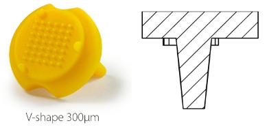
V Shape 300µ:
. Number of pins: 42
. Depth of the well: 1 mm
. Well bottom diameter: 300µm - Shape: circular
. Well upper side: 1mm*0.5mm - Shape: rectangular
V Shape 500µ:
. Number of pins: 42
. Shape of the pins: V
. Depth of the well: 1 mm
. Well bottom diameter: 500µm - Shape: circular
. Well upper side: 1mm*0.5mm - Shape: rectangular

Additional resources:
- Product overview
- More information on the Datasheet.
Results
hiPSCs cyst in alginate capsule in a V-shape well made with Stampwell
scale bar: 500µm

Image credit: Gaëlle Recher - Bordeaux
V-shape imprinted wells
Filled with a single cell-loaded alginate capsule: corresponding imaged with a stereomicroscope and stitched. Gel pad is made of 2% agarose. (Scale bar: 2mm)
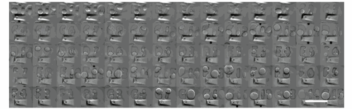
Image credit: Gaëlle Recher - Bordeaux
Protocol
How to use Stampwell:
- Read the full protocol: Stampwell Protocol
- Video protocol: How to use Stampwell

Protocol overview
1. Pour liquid agarose (or other hydrogel)
2. Place the stamp
3. Reticulate the hydrogel
4. Remove the stamp
FAQ
Find below advice & tips for Stampwell use:
Publications
Original publication:
Stampwell has been originally developed by Gaelle Recher and published in Scientific Reports:
Citations:
Merdrignac C, Clément AE, Montfort J, Murat F, Bobe J. auts2 Features and Expression Are Highly Conserved during Evolution Despite Different Evolutionary Fates Following Whole Genome Duplication. Cells. 2022 Aug 30;11(17):2694. doi: 10.3390/cells11172694. PMID: 36078102; PMCID: PMC9454499.
Sahai-Hernandez, P., Pouget, C., Eyal, S., Svoboda, O., Chacon, J., Grimm, L., Gjøen, T., & Traver, D. (2023). Dermomyotome-derived endothelial cells migrate to the dorsal aorta to support hematopoietic stem cell emergence. eLife, 12, e58300. https://doi.org/10.7554/eLife.58300
*본 상품은 오직 연구용으로만 사용 가능합니다. 인체 및 제품화에 사용하실 수 없습니다.

코아사이언스 coresciences Idylle France 프랑스 korea 한국 대리점 연구용 소모품 시약 세포생물학 cell imaging 세포 관찰 zebrafish imaging 제브라피쉬 이미징 관찰 UbiClear clearing kit for biological tissues SensiDeath - Ultra-sensitive human cell death assay a-Clipse - Chambers for luminescence imaging and photo/spectro-electrochemistry CrystalChip - Microfluidic chips for protein crystallization AgarSqueezer - agar-based cell compression device AgarSqueezer device AgarSqueezer - agar-based cell compression device SpheroTribe - all-in-one kit for reproducible spheroid/organoids Chitozen - Adhesive coverslips for bacteria imaging FakirSlide - Nanostructured glass coverslips for cell culture & isolated membranes Everspark - A long-lasting buffer for dSTORM GlowMito - Red fluorescent mitochondrial tracker BrightER - Endoplasmic reticulum probe for live imaging & flow cytometry ColorFlux - A visual indicator of bacterial efflux Stampwell V - 3D cell imaging Stampwell Z - Zebrafish imaging Stencell - Removable PDMS cell culture chambers AgarSqueezer Silicium Wafer SpheroRuler - Calibration beads for SMLM microscopy DNAbsolute - A column-free DNA extraction buffer Actiflash - A photoinducible protein activator Phimask - A phase mask to turn a 2D SMLM setup into a 3D image Stampwell U - 3D cell aggregation Luminicell Tracker™ - Cell Labelling Kit Luminicell Tracker™ - Vascular Labelling Kit
'실험.연구용 추천상품' 카테고리의 다른 글
- Total
- Today
- Yesterday
- 면역화학분석
- 콘드로이틴 황산 올리고당
- OLED
- 연속절편
- 오토클레이브백
- material science
- 파라핀 블록
- 후나코시
- gradient gel
- Sterlitech
- 바이오하자드백
- allview PAGE buffer
- 파라핀 블럭
- OPV
- filter
- 조직절편제작
- Funakoshi
- matrix-driven delivery pellet
- 미니멸균백
- 형광염색
- 건스터바이오텍
- 막필터
- solar cells
- 조직절편
- time release pellets
- 고수용성 콘드로이틴
- 코아사이언스
- 바이오헤저드백
- Coresciences
- 조직염색절편제작
| 일 | 월 | 화 | 수 | 목 | 금 | 토 |
|---|---|---|---|---|---|---|
| 1 | 2 | 3 | 4 | |||
| 5 | 6 | 7 | 8 | 9 | 10 | 11 |
| 12 | 13 | 14 | 15 | 16 | 17 | 18 |
| 19 | 20 | 21 | 22 | 23 | 24 | 25 |
| 26 | 27 | 28 | 29 | 30 | 31 |