

티스토리 뷰
[PROTEASOME/20S IMMUNOPROTEASOMES] 20S Immunoproteasome, human PBMC [SBB-PP0004]_South Bay Bio - 코아사이언스
코피디 2023. 7. 25. 14:5420S Immunoproteasome, human PBMC
Cat.# SBB-PP0004 / Size: 25 μg

Highly active protein complex that has been purified from human peripheral blood mononuclear cell (PBMC) . Immunoproteasome is recognized as a strong drug target for autoimmune disease and cancer. This immunoproteasome is supplied at >95% purity and experiments should be carried out at 20S immunoproteasome concentrations between 2-5 nM. [1][2][3][4]
Description
The immunoproteasome is structurally similar to constitutive 26S proteasome. The 20S core of immunoproteasome contains two outer rings composed of alpha subunits, and two internal 7-subunit containing rings each possessing 3 specific subunits responsible for proteasome catalytic activity. In immunoproteasome these subunits (ß1, ß2, ß5) are replaced by three inducible subunits: PSMB9, PSMB10, and PSMB8, (ß1i, ß2i, ß5i). These stress-induced subunits allow for the production of MHC-1 associating peptides, which are displayed as antigens on the cell surface. These displayed peptides can then be recognized by immune surveillance CD8 T-Cells. 20S Immunoproteasome is recognized as a strong drug target for autoimmune disease and cancer. This immunoproteasome is purified from human peripheral blood mononuclear cells and is supplied at >95% purity. Cells used as starting material tested negative for hepatitis B surface antigen, antibodies to hepatitis C virus, HIV type 1 antigens, and antibodies to HIV type 1 and 2. Immunoproteasome is commonly associated with the 19S, PA28 α/ß, or the PA28γ regulatory complexes. If choosing to omit PA28 during use, 20S must be chemically activated by addition of 0.035%SDS in final assay buffers.

Specifications
| Molecular Weight: | > 700 kDa |
| Purity: | > 95% SDS-PAGE |
| Storage Buffer: | 50 mM HEPES pH 7.5, 100 mM NaCl, 1 mM TCEP. |
| Storage | Store at −80°C after product arrival. Avoid multiple freeze / thaws. It is recommended to make multiple aliquots after the first thaw. |
Figure & Data
20S Immunoproteasome
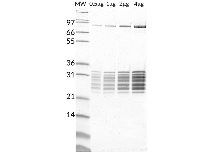
Figure 1. 20S Immunoproteasome, SDS-PAGE.
From left to right, increasing amounts of 20S Immunoproteasome loaded onto a 4-20% SDS-PAGE gel, stained with coomassie brilliant blue.
Immunoproteasome Activity

Figure 2. 20S Immunoproteasome vs. 20S Constitutive Proteasome Activity.
20S Immunoproteasome is most active against LLVY-AMC (SBB-PS0010), PAL-AMC (SBB-PS0007), and ANW-AMC (SBB-PS0009) substrates, representing physiologically relevant chemotrypsin-like, β1i, and β5i immunoproteasome activity respectively.
Citations & References
1) Wang J, Maldonado MA (Aug 2006). “The ubiquitin-proteasome system and its role in inflammatory and autoimmune diseases”. Cellular & Molecular Immunology. 3 (4): 255–61. PMID 16978533.
2) Murata S, Sasaki K, Kishimoto T, Niwa S, Hayashi H, Takahama Y, Tanaka K (Jun 2007). “Regulation of CD8+ T cell development by thymus-specific proteasomes”. Science. 316 (5829): 1349–53. doi:10.1126/science.1141915. PMID 17540904.
3) Cascio P, Hilton C, Kisselev AF, Rock KL, Goldberg AL (May 2001). “26S proteasomes and immunoproteasomes produce mainly N-extended versions of an antigenic peptide”. The EMBO Journal. 20 (10): 2357–66. doi:10.1093/emboj/20.10.2357. PMC 125470free to read. PMID 11350924.
4) Mallery DL, McEwan WA, Bidgood SR, Towers GJ, Johnson CM, James LC (Nov 2010). “Antibodies mediate intracellular immunity through tripartite motif-containing 21 (TRIM21)”. Proceedings of the National Academy of Sciences of the United States of America. 107 (46): 19985–19990. doi:10.1073/pnas.1014074107. PMC 2993423free to read. PMID 21045130.
* 본 상품은 오직 연구용으로만 사용 가능합니다. 인체 및 제품화에 사용하실 수 없습니다.

코아사이언스 coresciences South bay bio 한국 대리점 Ubiquitin proteasome system Real-time TR-FRET conjugation assays cryptases chemistry UB conjugation UB deconjugation C-terminal derivatives UBL derivatives inhibitors cryptate donors cryptate acceptors cryptate kits Di-ubiquitin tetra-ubiquitin immunoproteasomes 20S proteasomes substrates E1s E2s E3s ubiquitinated substrates DUBS
'실험.연구용 추천상품' 카테고리의 다른 글
- Total
- Today
- Yesterday
- 건스터바이오텍
- gradient gel
- 막필터
- 조직절편제작
- 형광염색
- 오토클레이브백
- 파라핀 블록
- 면역화학분석
- Sterlitech
- time release pellets
- solar cells
- 고수용성 콘드로이틴
- matrix-driven delivery pellet
- OLED
- 코아사이언스
- OPV
- 조직절편
- 미니멸균백
- 바이오하자드백
- 파라핀 블럭
- allview PAGE buffer
- filter
- 조직염색절편제작
- 연속절편
- 콘드로이틴 황산 올리고당
- 후나코시
- Coresciences
- 바이오헤저드백
- Funakoshi
- material science
| 일 | 월 | 화 | 수 | 목 | 금 | 토 |
|---|---|---|---|---|---|---|
| 1 | 2 | 3 | 4 | 5 | ||
| 6 | 7 | 8 | 9 | 10 | 11 | 12 |
| 13 | 14 | 15 | 16 | 17 | 18 | 19 |
| 20 | 21 | 22 | 23 | 24 | 25 | 26 |
| 27 | 28 | 29 | 30 | 31 |